
Pictures courtesy of Oral-B Laboratories.
 1. A healthy mouth has firm, pink gums that don't bleed. The gum is not attached to the tooth all the way to the top - there is a 1-2mm deep crevice around the gum line called the 'gingival sulcus', which can be measured with a special probe. Your toothbrush alone will not reach into the sulcus, so it is important to floss and brush twice daily.
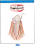
 2. Plaque causes the gum to become inflamed. Inflamed gums look puffy, red and swollen and they bleed easily. If left, the attachment of gum to tooth will deteriorate and the gingival sulcus will deepen. A measuring probe will show an abnormal 'pocket depth'. This condition can be treated with professional scaling and polishing and improving oral hygiene.
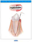
 3. As plaque accumulates it absorbs minerals from saliva and becomes hard. This rough, hard deposit is called 'calculus' or 'tartar'. This process can begin within just a few days of last cleaning your teeth. The physical damage caused to the gums along with increasing amounts of toxins released by the plaque eventually causes the bone that supports the tooth to shrink away. The gums begin to recede and the tooth may become loose. Treatment may involve professional deep scaling, root planing and polishing together with improved home care. Sometimes, gum surgery is needed, if so then specialist referral will be required.
 4. If the process is allowed to continue, advanced periodontitis ensues. The progress of this disease is usually completely pain-free. Patients who suffer from this condition are usually completely unaware of their bad breath, bleeding gums or discharge of pus from around their teeth. If left unchecked, bone loss becomes so severe that teeth become loose. Once the condition has progressed to this stage, teeth will usually need to be removed.
